New insights on atrial fibrillation through the integration of electrical rotors and fibrotic tissue analysis

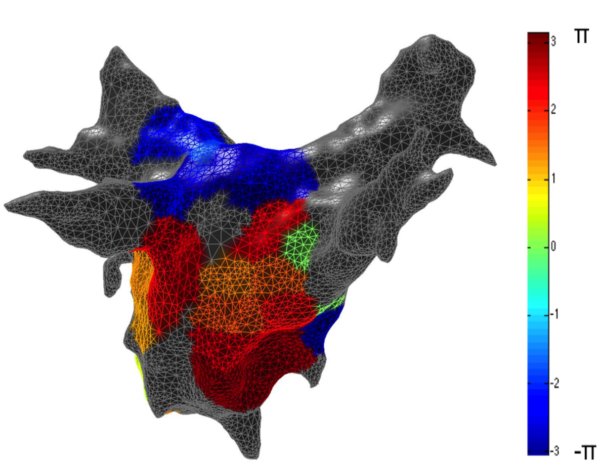
The PhD research project aims to study the link between atrial structural remodeling extent and electrical activations, focusing on the role of rotors and spatial relationship between them and atrial fibrosis. The project provides for development of 3D patient-specific left atrium (LA) model integrating anatomical and fibrosis information and a robust method to detect local atrial activation timings (LAATs) for phase map construction. In order to obtain the 3D anatomical model, magnetic resonance angiography (MRA) and delayed-enhanced MR imaging (DE-MRI) data are acquired and processed. The segmentation method of MRA data is obtained applying an edge-based level set approach guided by a phase-based edge detector. The analysis of the electrical patterns is based on the processing of the unipolar electrograms (EGM) acquired by a 64-pole basket catheter. The algorithm for identifying and quantifying the spatio-temporal organization of AF is based on the phase of the signals. From the integration of the information on the atrial fibrotic tissue extent and on the electrical activity is (will be?) possible analyze and study link between structural remodeling and electrical activations in AF.


